Introduction
One of the major concerns is soft tissue recession around implants that may appear over time (Adell et al. 1981; Grunder 2000) and this is an important issue not only for the patient but also for the clinician.
Today one of the first considerations for implant therapy is to achieve aesthetic results that are in harmony with the surrounding tissues.
Although this is a very familiar finding in our daily practice, literature is lacking regarding approaches to solve this problem of recessions involving implants predictably (Roccuzzo et al. 2002; Mareque-Bueno 2011; Zucchelli et al.2012).
Soft Tissue Recession Around Implants
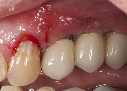
A 62 year old lady attended for a review appointment, her main complaint was the recession around an implant in the #14 implant position. The patient is fit and healthy and a nonsmoker. Let’s consider the causes that could lead to this recession. I scored the following causes with 0 if it is very improbable to be the cause of the recession, one maybe, and two whereas it is probably the main cause:
- Mucosal quality (keratinized vs. Non-keratinized)
This is improbable to be the main cause. The amount of keratinized tissue is more than 1 mm and this is unlikely to be a cause so the score for this one is 0.
- Mucosal attachment (mobile vs. Non-mobile)
There was enough attached mucosa and we should discard this cause. Score 0.
- Mucosal thickness
Very thin mucosa was found. This is probably one of the main causes. Score 2.
- Facial bone crest level and thickness
During the peri-implant probing depth (PPD) at the buccal aspect was +/- 6 mm so we should give this a score 2.
- Implant fixture angle
The angulation of the abutment was correct from the prosthetic and biological point of view. Score 0.
- Interproximal bone crest level
Both interproximal peaks of the bone crest were well preserved. Score 0.
- Level of first bone to implant contact
As I previously mentioned, the depth of probing was 6 mm. Score 2.
The platform depth was above the bone crest. Score 0.
- Micro and macrostructure of the implant neck.
The microstructure is micro threads and would not influence soft tissue recession. Score 0.
- Implant-abutment and prosthesis connection
I used an intermediate abutment (Aurea, Phibo, Spain), during the impressions to avoid recession caused by the connection and disconnection of the abutments. Score 0.
- Surgical technique
The surgical procedure was uneventful. 2 years ago a sinus floor augmentation was performed allowed to consolidate before implants were placed. Score 0.
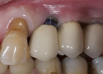
The total score was 6 out of 22 where over 10 we should consider that maybe the treatment option should be to remove the implant and site form the area to avoid recession????
After reviewing all this, I made a decision based on the score of 2. We have a thin facial bone crest and thin mucosa, and the level of the first bone to implant contact was low, so we had to solve two points.
- Bone thickness and first implant-bone contact: The approach here should involve GBR with a bone substitution and a membrane
- Thin soft tissue: At this point, the only option was to perform a soft tissue augmentation.
Considering the two approaches described previously, I decided to perform a guided bone regeneration simultaneously to the soft tissue augmentation.
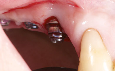
Surgery to Solve Soft Tissue Recession
The first step was to remove the fixed prosthesis. This will allow us to perform a coronally advanced flap and will facilitate both advanced coronally flap surgery and connective tissue harvesting surgery from the palatal.
First of all, the connective tissue graft was harvested from the palatal. 4/0 PTFE sutures were used to suture the wound.
A flap with beveled distal and mesial vertical releasing incisions was raised, avoiding the canine and the adjacent implant. Several incisions were performed apically in the periosteum so the flap could be mobilized coronally and cover the recession.
GBR was performed utilizing Bio-Oss (Geistlich, Switzerland) mixed the autologous bone (50/50) and a collagen membrane Creos (Nobel Biocare). The membrane was cut in two pieces and used to cover the biomaterial, one was placed vertically and the other horizontally so it can stabilize the biomaterial.
The connective tissue was then sutured to the implant neck with 5/0 monofilament sutures and covered with the flap.
Antibiotic amoxicillin 500 mg and anti-inflammatories were prescribed to the patient for 5 days.

Conclusion
After 15 days of healing the patient came to the office and an improvement in the soft tissue level could be observed. Long term observation is needed to confirm the improvement of the soft tissue thickness and the partial coverage of the recession.
Although soft tissue management around peri-implant tissues is not always predictable, but as in teeth some approaches can be performed to solve soft tissue recessions around implants such as a coronally repositioned flap with simultaneous connective tissue graft.
